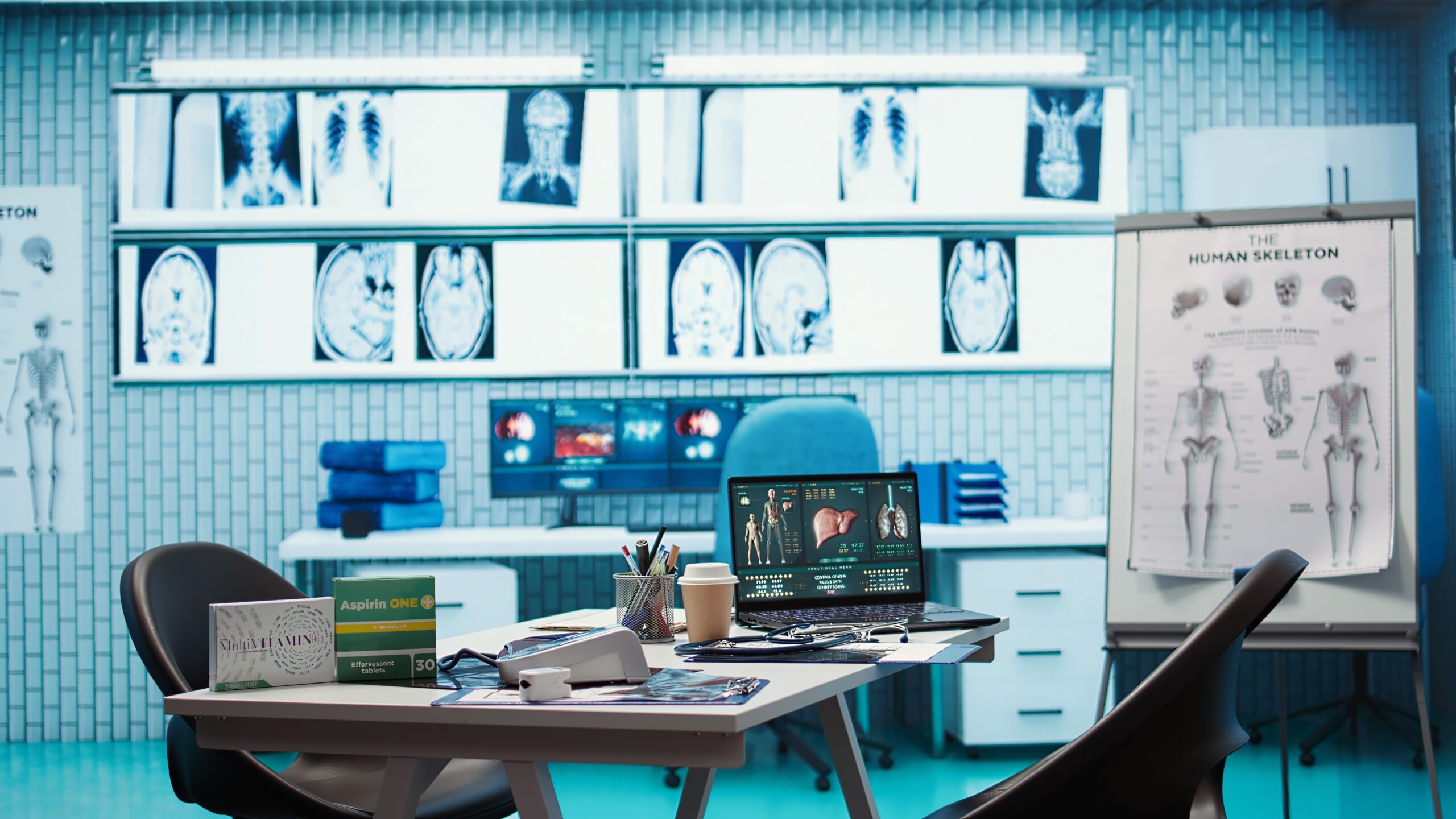
In the world of modern healthcare, Diag Image—short for diagnostic imaging—plays a pivotal role. It refers to the technologies and methods used to create visual representations of the inside of the body, helping doctors diagnose, monitor, and treat a wide range of medical conditions.
Over the decades, diagnostic imaging has evolved from basic X-rays to highly advanced tools like MRI, CT scans, and PET imaging. These innovations have transformed the way physicians understand, visualize, and interact with the human body—offering faster, safer, and more accurate diagnostics than ever before.
What is Diag Image?
The term Diag Image encompasses a collection of imaging modalities used to capture images of organs, bones, tissues, and even cellular processes within the body. This includes traditional methods like X-rays and ultrasounds, as well as more sophisticated systems such as MRI and PET scans.
Notably, the core advantage of diagnostic imaging lies in its non-invasive nature. Rather than performing exploratory surgery, doctors can use imaging to pinpoint the location, size, and behavior of abnormalities—often in real time and without discomfort to the patient.
The Role of Diag Image in Healthcare
Diagnostic imaging is integral to nearly every medical specialty. Whether diagnosing a fractured bone, detecting a tumor, or monitoring heart health, imaging provides the clarity clinicians need. It reduces uncertainty, guides treatment decisions, and ultimately improves patient outcomes.
Additionally, Diag Image supports preventive medicine by identifying early signs of disease—long before symptoms appear. This proactive capability empowers healthcare providers to intervene sooner, often improving the chances of a successful outcome.
Historical Evolution of Diagnostic Imaging
Diag Image has a rich history dating back to the discovery of X-rays in 1895 by Wilhelm Roentgen. This groundbreaking moment marked the beginning of a revolution in medical diagnostics. Over time, advancements like ultrasound (1950s), CT scans (1970s), and MRI (1980s) pushed the boundaries of what imaging could achieve.
Today, the field is experiencing another leap forward with the integration of digital imaging, AI-based interpretation, and portable devices that bring diagnostics to the bedside.
Key Types of Diag Image Technology
X-ray Imaging
X-rays use electromagnetic radiation to capture images of dense tissues, such as bones and teeth. They are quick, cost-effective, and widely available, making them ideal for detecting fractures, lung infections, and dental issues.
CT Scans (Computed Tomography)
CT scans combine multiple X-ray images taken from different angles to produce cross-sectional views of the body. They offer more detail than standard X-rays and are commonly used to assess trauma, internal bleeding, and cancers.
MRI (Magnetic Resonance Imaging)
MRI uses magnetic fields and radio waves to produce detailed images of soft tissues, including the brain, muscles, and ligaments. Since it doesn’t involve radiation, it’s considered safer for repeated use—particularly in neurology and musculoskeletal imaging.
Ultrasound
Ultrasound emits high-frequency sound waves that bounce off body tissues to create real-time images. It is widely used in obstetrics, cardiology, and abdominal imaging. The technology is safe, portable, and non-invasive.
PET (Positron Emission Tomography)
PET scans involve injecting a small amount of radioactive tracer into the body. This allows clinicians to observe metabolic activity, which is especially useful in cancer diagnosis, neurological disorders, and evaluating organ function.
Hybrid Imaging
Combining imaging modalities—like PET/CT or PET/MRI—enhances diagnostic accuracy by merging structural and functional data into one comprehensive image.
Applications of Diag Image in Clinical Practice
Emergency Medicine
When seconds count, Diag Image provides life-saving clarity. Tools like CT and ultrasound help identify internal injuries, strokes, and blockages swiftly, allowing for immediate treatment.
Oncology
Diag Image is critical in cancer detection, staging, and treatment monitoring. By visualizing tumors and surrounding structures, doctors can target therapies more effectively and monitor their success.
Neurology
MRI and CT scans are invaluable in assessing brain injuries, strokes, multiple sclerosis, and other neurological disorders. These tools offer unparalleled detail of the central nervous system.
Orthopedics
In bone and joint disorders, diagnostic imaging aids in identifying fractures, degenerative conditions, and ligament damage. It also assists surgeons in planning procedures and evaluating recovery.
Cardiology
Echocardiograms, cardiac CT, and MRI provide insights into heart structure and function. These imaging methods are essential for diagnosing heart disease, valve disorders, and vascular abnormalities.
Benefits of Diag Image
- Non-Invasive and Painless: Most imaging techniques avoid incisions or invasive procedures, making them safer and more comfortable.
- High Accuracy: Diag Image enhances diagnostic precision, allowing doctors to differentiate between similar conditions.
- Faster Diagnosis: With real-time results available in many cases, imaging reduces delays in treatment.
- Improved Monitoring: Repeated imaging enables doctors to track the progression or regression of diseases over time.
- Cost-Effective: By eliminating unnecessary surgeries, imaging reduces overall healthcare costs.
Recent Innovations in Diag Image
AI and Machine Learning
Artificial intelligence is transforming Diag Image by assisting radiologists with pattern recognition, anomaly detection, and image classification. AI can prioritize urgent findings, improve accuracy, and reduce workload.
Portable Imaging Devices
Handheld ultrasound machines and portable X-ray systems are making it possible to bring diagnostic tools to the patient’s bedside—or even to remote villages.
Digital and 3D Imaging
New digital platforms offer sharper images and better storage. 3D imaging allows for virtual reconstruction of organs and structures, supporting surgical planning and educational visualization.
Low Radiation Technologies
Emerging systems now use less radiation without compromising image quality. These innovations are particularly valuable in pediatric imaging and chronic illness management.
Challenges and Considerations
While Diag Image offers remarkable benefits, it’s not without limitations:
- Radiation Exposure: Repeated exposure to X-rays and CT scans must be carefully managed.
- Interpretation Errors: Even skilled radiologists can miss subtle signs; human error remains a concern.
- Access and Affordability: In many parts of the world, diagnostic equipment is scarce or prohibitively expensive.
- Data Privacy: Digital imaging must comply with strict privacy regulations to protect patient information.
The Future of Diag Image
Diagnostic imaging is entering a new era—one where AI, genomics, and remote diagnostics converge. Soon, we may see:
- AI-Powered Triage Systems that prioritize scans based on urgency
- Tele-Imaging platforms allowing specialists to interpret scans from anywhere in the world
- Integration with Genomics to create full-spectrum patient profiles
- New Imaging Modalities capable of visualizing single cells or tracking treatments in real-time
As technology advances, Diag Image will not only diagnose disease but predict it—shaping the future of precision medicine.
FAQs
What is Diag Image used for?
It’s used to non-invasively visualize internal body structures for diagnosis and treatment planning.
Is diagnostic imaging safe?
Yes. Most imaging techniques are safe, though some involve minimal radiation. Safety protocols are strictly followed.
Do you need preparation before imaging?
Sometimes. Preparation may include fasting, removing metal, or taking contrast agents, depending on the test.
How long do results take?
Basic tests like X-rays may be available immediately. MRIs or PET scans can take several hours to a few days.
Can imaging detect all diseases?
Not all. While powerful, diagnostic imaging is often combined with labs and physical exams for accurate diagnosis.
What’s the role of AI in imaging?
AI helps detect patterns, speeds up interpretation, and supports radiologists in making more accurate diagnoses.
Why Diag Image Is the Backbone of Modern Diagnostics
In today’s healthcare landscape, Diag Image stands out as a transformative force. It equips physicians with the clarity needed to diagnose earlier, treat smarter, and monitor progress more effectively. Far from being just a snapshot of the body, imaging is becoming a dynamic, intelligent system—one that learns, adapts, and enhances the entire medical journey.
Looking forward, the fusion of AI, digital health, and imaging technology promises to elevate patient care to unprecedented heights. Diag Image isn’t just part of modern medicine—it’s defining it.